Information center / Breast cancer / Cancer / Monthly Spotlight / Prevention / Radiology / Science / VM Med / Women's health
Breast Cancer: Early Detection & What Happens At Each Stage

(Tina Dawn/ VM Med) — Breast cancer is the most common cancer in Canadian women, and the second leading cause of cancer-related deaths. One in eight women will be diagnosed with breast cancer in their lifetime. Knowing this, preventative measures and vigilance should always be a top priority and a fundamental part of women’s healthcare.
The most common symptoms of breast cancer are:
· A new lump in the breast or armpit
· Thickening or swelling of part of the breast
· Irritation or dimpling of the breast skin
· Redness or discharge in the nipple area
· Fatigue or constant cough
It’s important to remember, however, that many women with breast cancer have no symptoms. This is why regular breast cancer screening and routine mammograms are so important.
Mammograms can help find both cancerous (malignant) and non-cancerous (benign) tumours in the breast. Early detection of breast cancer can lead to more successful treatment and a higher chance of being cured.
The Sooner, The Better

There are 5 stages of breast cancer – stage 0 followed by stages 1 to 4. Generally, the higher the stage number, the larger the cancer is or the more the cancer has spread.
Ideally, you want to detect it in early stage breast cancer while the tumour is still small, confined in the breast tissue, and has not had the chance to spread to the lymph nodes.
Stage 0 breast cancer, also referred to as breast cancer in situ means that the cancer cells have not spread at all and are only in the duct where they started.
Stage 1 breast cancer usually means the tumour is approximately 20 mm or smaller in diameter and there has been none or very little spread of cancer to the lymph nodes. The prognosis for Stage 1 breast cancer is excellent with the 5-year relative survival rate at 99 percent.
Stage 2 breast cancer is when the tumour is larger than 50 mm but has not spread to any lymph nodes.
Stage 3 breast cancer is when the tumour is larger than 50 mm, has spread to a few lymph nodes but has not spread to the rest of the body.
Stage 4 is when the cancer has spread to other parts of the body, also known as metastatic breast cancer.
The Canadian Cancer Society does a thorough job of describing in further detail all stages of breast cancer and why early detection is best.
Treatment for breast cancer depends on what stage is diagnosed. Early stage breast cancers are often treated with surgery and radiation, while later stages usually involve a combination of surgery, chemotherapy, radiation, or hormone therapy. The approach depends on each individual and the cancer type.
Early-stage Mammogram vs Early-stage Ultrasound
A mammogram is always the best way to detect early stage breast cancer. It is the primary screening tool for breast cancer. A breast ultrasound, on the other hand, is most often done to further clarify if a problem found by a mammogram or physical exam of the breast is a cyst filled with fluid or in fact a solid tumour. An ultrasound is basically the test that follows when a change has been seen on a mammogram. It’s not a replacement screening tool for a mammogram.
When it comes to early detection of breast cancer, a mammogram is the diagnostic test that will provide a more complete photo of the breast, identifying any lumps, changes, or asymmetry that may be causes of concern for your doctor.
A breast ultrasound, on the other hand, provides more detailed imaging and information about a particular area of your breast and allows your doctor to further look at any breast abnormalities.

What to Expect at a Mammogram Appointment
A mammogram is a pretty straightforward and uncomplicated test that doesn’t require much in terms of preparation.
The Canadian Cancer Society recommends you avoid using anything under your arms or on your breasts that can affect the accuracy of the x-ray the day of your mammogram. Deodorant, antiperspirant, lotions, creams, powders or perfumes should be avoided and may need to be wiped off before your test.
It’s helpful to wear a top and bra that are easy to take off. Remove necklaces and other jewellery that might get in the way during the test.
The test itself is described as mildly uncomfortable by most patients. If you’re worried about pain, it’s best to avoid getting a mammogram the week before or after your menstrual period when your breasts are tender and occasionally even sore. Some experts also recommend having less caffeine for 2 weeks before your mammogram, which can also help with pain management. You can also take painkillers like Advil or Tylenol an hour before your x-ray.
You’ll be asked to undress from the waist up. Then, during the mammogram, the radiology technologist will position your breast on a special x-ray machine. The breast is placed between 2 plastic plates. The plates are slowly pressed together to flatten the breast and restrict any motion, allowing for images to be as clear as possible. Usually, four images are taken during a mammogram, two of each breast from two different angles.
If at any time you experience extreme discomfort, let the radiology technologist know to adjust the pressure. The entire appointment usually lasts about 15 minutes.
There are 2 kinds of mammograms. A screening mammogram that is done to screen women with no signs of breast cancer. Diagnostic mammograms help diagnose women with signs of breast cancer.
Why a Breast Cancer Ultrasound?
Women with dense breasts may benefit from getting a breast ultrasound or additional screening, since the density of their breast tissue makes it difficult for a mammogram to detect signs of cancer.
Women with breast implants may require additional screening or extra images since the implants may sometimes affect the results or make it difficult for the radiology technician to see the breast tissue and detect signs of cancer.
A breast cancer ultrasound is similar in duration and nature to a mammogram. You should avoid putting lotion or powder on your breasts on test day. You’ll be asked to take off any jewelry and clothing from the waist up.
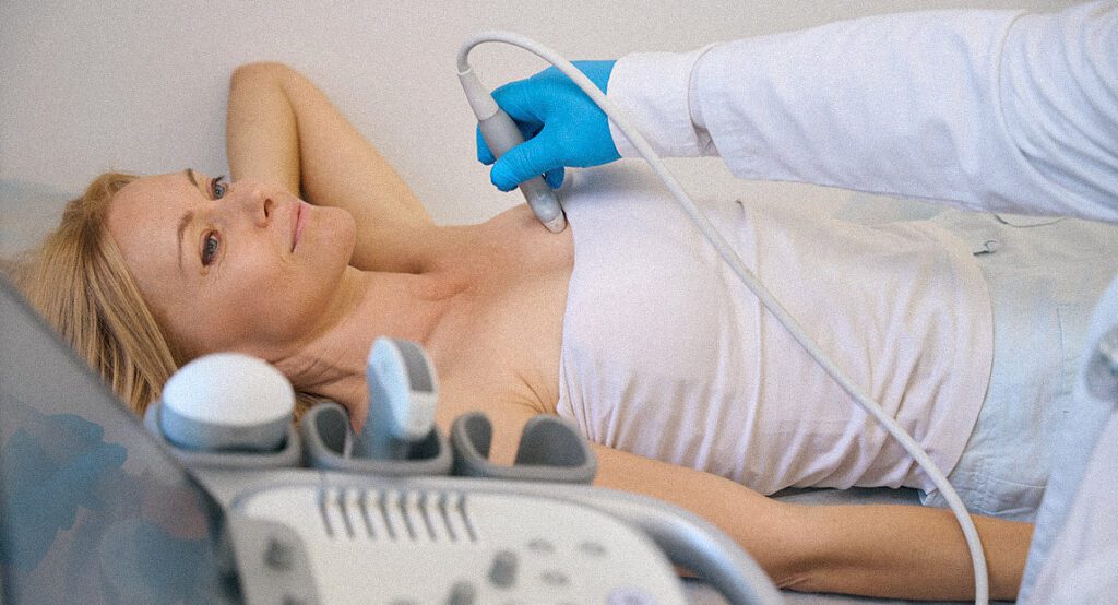
You’ll lie on your back and occasionally on your side on an exam table. You’ll be asked to raise your arm above your head and the technologist will put a clear gel on your skin over the breast area. The technologist will press the transducer (a device that produces sound waves that bounce off body tissues and make echoes) against your skin and slide it over the area being studied. Once the test is concluded, the gel will be wiped off.
Ultimately, what matters is detecting and treating breast cancer as early as possible. This increases both the odds of successful treatment and better prognosis and decreases the need for more invasive treatments. Monthly breast self-exams and regular clinical breast exams and mammograms are a key component of your healthcare.
VM Med Breast Center
VM Med’s Breast Center, the largest private breast center in Canada, is dedicated to taking care of every aspect of our patients’ breast health needs. Our team of experts assesses each individual breast cancer case and works with you to create a personalized course of treatment that prioritizes your health and comfort throughout this process. Our services include breast imaging, breast biopsy, breast surgery, breast cancer staging, breast oncology, and breast cancer genetic testing.
For more information on your screening, you can read our extensive archive of VM Med blogs, including Young Women Shouldn’t be Non-Chalant About Breast Cancer, or How to Perform a Breast Self-Examination.
Still have questions? Book a consultation with our experts.
Categories